
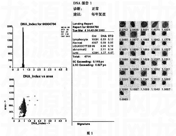
 总结各种子宫颈癌筛查方法的经验基础上,一方面,本研究采用子宫颈刷取材和国产化的液基薄层制片技术,同时使用扫描和诊断过程全自动化的DNA倍体分析系统进行筛查。 另一方面对基层妇女进行宣传教育,使她们了解子宫颈癌早防早治的意义,受到了广大育龄妇女的欢迎并积极参与。经过大力宣传,主动参加全自动DNA分析系统进行子宫颈癌普查的基层妇女人数增长。实践证实用全自动DNA倍体分析系统进行子宫颈癌普查有效地解决了在基层开展大规模子宫颈癌普查缺乏应有的技术和欠缺有经验细胞学医生的矛盾。同时该方法敏感度较高,成本效益合理,应作为一种适合于基层大规模妇女普查的子宫颈癌筛查方法。
【参考文献】
[1]Liu S, Semenciw R, Probert A, et al. Cervical cancer in Canada: changing patterns in incidence and mortality. Int J Gynecol Cancer,2001, 11: 24-31.
[2]Anderson GH, Boyes DA,Benedet JL, et al .Organisation and results of the cervical cytology screening programme in British Columbia, 1955-1985. Br Med J ,1988,296: 975-978.
[3] Guidozzi F. Screening for cervical cancer.Obstet Gynecol Surv, 1996,51:696-701.
[4] Hutchinson M, Fertitta L, Goldbaum B, et al. Comparison of their ability to sample abnormal cells for cervical smears. J Reprod Med , 1991, 36 :581-586.
[5] Laverty CR, Farnsworth A, Thurloe JK, et al. The importance of the cell sample in cervical cytology: a controlled trail of a new sampling device. Med J Aust , 1989,150 :432-436.
[6] Doornewaard H , van der Graaf Y. Contribution of the cytobrush to determining cellular composition of cervical smears. J Clin Pathol , 1990,43 : 393-396.
[7] McCord ML, Stovall TG, Meric JL, et al. Cervical cytology: a randomized comparison of four sampling methods. Am J Obstet Cynecol , 1992,166 :1772-1779.
[8] Stenkvist B , Soderstrom J. Reasons for cervical cancer despite extensive screening. J Med Screen , 1996,3 : 204-207.
[9] Kohlberger PD, Stani J, Gitsch G, et al . Comparative evaluation of seven cell collection devices for cervical smears. Acta Cytol, 1999,43:1023-1026.
[10]Jarvi K. Cervex brush versus vaginal-cervical-endocervical (VCE) triple smear techniques in cervical sampling. Cytopathology , 1997,8 :282-288.
[11]Vooijs GP. Endocervical brush device. Lancet, 1989,1 : 784.
[12]Waddell CA, Rollason TP, Amarill JM, et al. The Cervex: an ectocervical brush sampler. Cytopathology, 1990, 1: 171-181.
[13]Lee KR, Ashfaq R, Birdsong GG, et al . Comparison of conventional Papanicolaou smears and a fluid-based, thin-layer system for cervical cancer screening. Obstet Gynecol, 1997,90:278-284.
[14]Wilbur DC, Cibas ES, Merritt S, et al. ThinPrep Processor. Clinic trials demonstrate an increased detection rate of abnormal cervical cytologic specimens. Am J Clin Pathol, 1994,101 :209-214.
[15]Tezcan A, Garner DM, Lam P, et al . Analysis of thionin, gallocyanin and hematoxylin for automated quantitative image cytometry of cervical samples. 8th Annual Meeting, Clinical Applications of Cytometry, 1993, pp:15-18.
[16]Palcic B, Garner DM, MacAulay CE, et al. Oncometrics Imaging Corporation and Xillix Technologies Corporation. Use of the Cyto-Savant in quantitative cytology. Acta Cytol , 1996,40 : 67-72.
[17]Doudkine A, MacAulay C, Poulin N, et al. Nuclear texture measurements in image cytometry, Pathologica, 1995,87 :286-299.
[18]Bcking A, Adler CP, Common HD, et al. Algorithm for DNA cytophotometric diagnosis and grading of m alignancy. Anal Quant Cytol Histol. 1984;6:1-7.
[19] Bcking A, Hilgarth M, Auffermann W, et al. DNA-cytometric diagnosis of prospective m alignancy in borderline lesions of the uterine cervix. Acta Cytol. 1986;30:608-615.
[20]Grote HJ, Friedrichs N, Pomjanski N, et al. Prognostic significance of DNA cytometry in carcinoma of the uterine cervix FIGO Stage IB and II. Anal Cell Pathol. 2001;23:97-105.
[21]Fu YW, Reagen JW, Fu AS, et al. Adenocarcinoma and mixed carcinoma of the uterine cervix. 2. Prognostic value of nuclear DNA analylsis. Cancer. 1982;49:2572-2577.
[22]Chatelain R, Schunck T, Schindler EM, et al. Diagnosis of prospective m alignacy in koilocytic dysplasia of the cervix with DNAcytometry. J Reprod Med. 1989;34:505-510.
[23]Kashyap V, Das DK, Luthra UK. Microphotometric nuclear DNA analysis in cervical dysplasi of the uterine cervix:its relation to the progression to m alignancy and regression to normal. Neoplasma. 1990;37:487-500.
[24]Bollmann R, bocking A. Prognostic validity of DNA-imaging-cytometry in cervical dysplasias. Verh Dtsch Ges Path. 1996;80:557.
[25]Hering B, Horn LC, Nenning H, et al. Predictive value of DNA cytometry in CIN 1 and 2. Image analysis of 193 cases. Anal Quant Cytol Histol. 200;22:333-337.
[26]Torsten W. Remmerbach, Horst Weidenbach, Natalja Pomjanski, Kristinae Knops, Stefanie Mathes, Alexander Hemprichc and Alfred Bcking. Cytologic and DNA-cytometric early diagnosis of oral cancer. Analytical Cellular Pathology 22 (2001) 211-221.
[27]Azua J. Romeo P. Morales M et al. DNA quantification as a prognostic factor in gastric adenocarcinoma. Anal Quant Cytol Histol 1998:20:221-4.
[28] Dey P. Luthra UK. Prasad A et al. Cytologic grading and DNA image cytometry of breast carcinoma on fine needle aspiration cytology smears. Anal Quant Cytol Histol 1999:21;17-20.
[29]Murty UV, Mitra AB, Das BC, et al. Chromosomal Phenotypes in patients with precancerous lesions of the uterine cervix progressed to cancer during follow up. Oncology. 1988;45:384-388.
[30]Norming U, Tribukait T, Gustafson H, et al. Deoxyribonucleic acid profile and tumor progression in primary carcinoma in situ of the bladder: a study of 3 patients with Grade 3 lesions. J Urol. 1992;147:11-15.